Book Appointment

Dental X-Rays & Advanced Imaging in Pleasant Prairie

— see what the naked eye can’t
Dental imaging helps us identify hidden oral health concerns before they become more serious and allows us to plan treatment with greater accuracy. At Old Oaks Dental, we use modern digital X-rays and advanced imaging technology to capture clear, detailed images while keeping radiation exposure as low as reasonably possible.

Modern Digital X-rays use up to 90% less radiation than film

The majority of dental problems are invisible without imaging

Standard visual screenings only reveal surface enamel health. Critical underlying dynamic concerns — including deep decay patterns hiding between tight teeth, early jawbone reabsorption tracks, hidden root abscesses, impacted third molars, cysts, and tumor paths — stay completely concealed beneath structural surfaces.

Dental radiography isn’t an elective operational add-on; it is an irreplaceable baseline patient protection mechanism. We frame diagnostics strictly around preventative security, never revenue generation fields.

Imaging Cadence Framework

Bitewing Radiographs: Every 1–2 years (Routine Check)

Full Mouth Series (FMX): Every 3–5 years (New Patient Baseline)
Panoramic Screening: As needed for implants, wisdom teeth, ortho
3D Cone Beam (CBCT): Reserved for implant design & complex paths

Full Scope Checklist

What we assess during your comprehensive evaluation

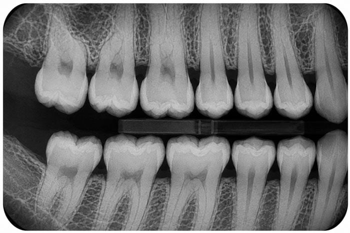
Bitewing dental X-ray showing the upper and lower back teeth
Every individual tooth

Tactile examination checking for decay, fracture lines, enamel attrition wear, and aging restoration integrity status fields.

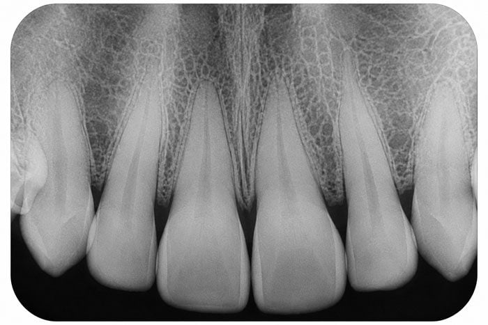
Periapical dental X-ray showing the roots of the upper teeth
Periapical X-rays

Displays an entire singular tooth profile from top crown down to the root tip anchor bone density. Used to verify root canal infections or absolute fracture paths.

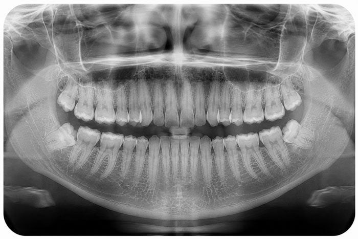
Panoramic dental X-ray of the upper and lower jaw
Panoramic X-ray

Generates a single wide-view flat map layout of the entire maxilla and mandible arches, jaw joints (TMJ), and sinus floors. Vital for wisdom teeth mapping.

Full-mouth series of dental X-rays with a panoramic view at the centre
Full Mouth Series (FMX)

A comprehensive structural set of 14–20 individual localized digital sensors. Captures the complete baseline diagnostic grid for incoming guest charts.

Cone beam CT scan images showing the jaw from several angles
CBCT (3D Cone Beam)

Computes a highly accurate 3D volumetric model of bone thickness, nerve networks, and vascular pathways. Critical for zero-compromise surgical implant plotting.

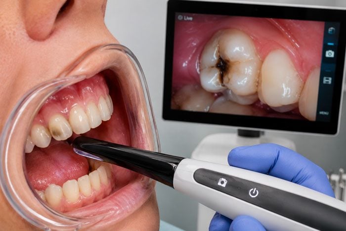
An intraoral camera in use, with the image of the tooth shown on a screen
Intraoral Camera

A pen-sized micro-wand lens displaying high-definition live color video of internal tooth surface fractures or leakage margins right onto your chairside monitor.

Uncompromising Safety Protocol

Our proactive approach to radiation protection

Up to 90% less exposure

Modern high-speed digital sensors capture structural diagnostic layouts instantly with up to 90% less required energy than historical analog film sheets. A routine check exposes you to less background radiation than a brief domestic airline flight.

Lead apron barriers

We universally implement heavily lined lead aprons paired with built-in thyroid shielding collars for all basic radiograph procedures, establishing an absolute physical line of secondary environmental tissue protection.

Only when clinically necessary

We do not support automatic calendar-based baseline imaging templates. We strictly align tracking intervals with custom ADA risk guidelines; each diagnostic order is mapped independently to your individual biology.
“We understand many families navigate valid questions regarding cumulative radiation exposures from medical and dental diagnostics. Here are the clear, scientific safety boundaries we enforce daily.”
Precision Diagnostics That Catch What Others Miss

Cutting-edge technology

From instant ultra-high-definition digital bite sensors to advanced low-dose CBCT 3D scanning, our hardware infrastructure minimizes capture delays and maximizes data clarity.

Chairside image sharing

We present images clearly on our large high-res monitors, mapping out anatomy in plain, plain-English terms so you maintain full diagnostic transparency alongside our team.

Secure digital records

All clinical diagnostic images load into protected storage loops instantly. This lets us run overlay comparisons year-over-year to catch subtle, silent bone pattern losses early.

Real Patient Feedback

True transformation stories highlighting our compassionate, relation-first hygiene protocols.
Google reviews

Trusted by local families

4.9 ★★★★★ Google
Maria Zavala
Maria Zavala1 month ago
★★★★★

Everyone in the office are truly wonderful people! Was treated with respect and will definitely…

Nadine Burge
Nadine Burge5 months ago
★★★★★

There’s no better dental office than old oaks. Everyone is so nice and knowledgeable. Wish…

Shari Short
Shari Short4 weeks ago
★★★★★

Bridget was excellent and very detailed in my procedure and cleaning!

Darcey Stringer
Darcey Stringer5 months ago
★★★★★

Dr. Dana, Melissa, and the rest of the staff are great! Definitely the first time…

Kaylyn Eagney
Kaylyn Eagney5 months ago
★★★★★

Such a friendly, happy staff that makes you feel super comfortable and informed with the…

Gloria Evangelista
Gloria Evangelista5 months ago
★★★★★

Thanks to Dr. Dana for fixing my tooth

Michelle Long
Michelle Long1 month ago
★★★★★

Bridget is amazing, goes over everything in detail, gives clear answers to my questions and…

Russell Fisher
Russell Fisher3 months ago
★★★★★

An awesome experience, where the staff made me feel incredibly comfortable. Second to none service.

David Jackson
David Jackson5 months ago
★★★★★

Excellent job! All women workers and dentists…. been my favorite for years.

Charles Christensen
Charles Christensen5 months ago
★★★★★

Excellent Job. Very nice people working there.

Bob Trapp
Bob Trapp2 months ago
★★★★★

A very friendly and competent staff👍👍😀

Jackie Allen
Jackie Allen4 months ago
★★★★★

This is my second visit to old oak dentistry. Since the retirement of Dr. Dan….

Frequently Asked Diagnostic Questions

For healthy adult recall patients, localized digital bitewing checks are suggested every 12 to 24 months. New patients typically require a full mouth series or structural panoramic scan to anchor an accurate, baseline tracking metric.

Yes. Because digital capture arrays cut down output thresholds by up to 90% compared to legacy film layers, radiation exposure is remarkably nominal. When combined with our protective lead apron layering, the procedure is verified safe across children and expectant mothers.

While we always honor your ultimate treatment autonomy, declining necessary diagnostic imaging completely compromises our clinical capacity to screen for sub-surface path trends or safely clear teeth for clinical adjustments. We document your parameters meticulously and work within safety parameters.

Cone Beam Computed Tomography rotates a specialized sensor array to capture a precise 3D rendering of bone depth, neural paths, and sinus layouts. It is universally required for accurate surgical implant design, complex wisdom extractions, and advanced airway evaluations.

Standard basic diagnostic images like bitewings or baseline panoramic scans are almost universally covered fully (at 80% to 100%) within preventive insurance allowances. High-tier surgical 3D CBCT scans can occasionally necessitate pre-authorization.

Related services

Comprehensive oral exam

Advanced diagnostic radiographs are reviewed and evaluated as a core piece of your complete baseline head and mouth health assessment.

Dental implants

High-resolution 3D CBCT mapping generates accurate structural data fields, securing safe, predictable prosthetic implant placements.

Orthodontics & alignment

Panoramic and cephalometric radiography workflows map bone scaling indexes, tracking path metrics ahead of clear aligner placement paths.

Open for Appointments

Schedule your visit with our friendly dental team today.